


F4/80 Monoclonal Antibody (BM8), Super Bright™ 436, eBioscience™
View Cart or Continue shopping.
Description
PRODUCT DETAILS
Host: Rat
Isotype: IgG2a, kappa
Clonality: Monoclonal
Clone: BM8
Format: Super Bright™ 436
Reactivity: Mouse
Application: Flow Cytometry
Tested Dilution: 1.0 µg/test
Concentration: 0.2 mg/mL
Storage: 4° C, store in dark, DO NOT FREEZE!
Formulation: PBS, pH 7.2, containing 0.09% sodium azide
Purification: Affinity chromatography
Data Sheet: TDS
Specific Information
Description: The BM8 monoclonal antibody reacts with mouse F4/80 antigen, an approximately 160 kDa surface receptor. It belongs to the EGF-TM7 family of proteins. As such it contains seven EGF-like domains on its extracellular N-terminus, seven transmembrane spanning sequences, and an intracellular C-terminal domain showing homology to other TM7 superfamily members. The F4/80 antigen is expressed by a majority of mature macrophages and is one of the best markers for this population of cells. However, other cell types, such as peritoneal eosinophils, Langerhans cells, and some other dendritic cell subtypes, have been reported to express this antigen as well. Expression of F4/80 commences during early myeloid development in vivo and can be upregulated on BM cells stimulated in vitro with M-CSF. Some populations of macrophages, especially in the lymphoid microenvironment, may be devoid of F4/80.
Applications Reported: This BM8 antibody has been reported for use in flow cytometric analysis.
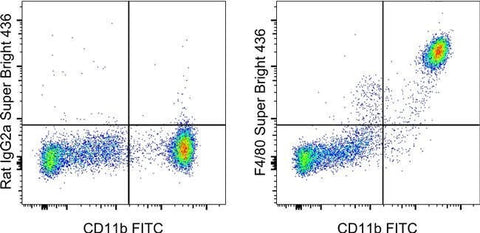
Applications Tested: This BM8 antibody has been tested by flow cytometric analysis of resident peritoneal macrophages. This may be used at less than or equal to 1.0 µg per test. A test is defined as the amount (µg) of antibody that will stain a cell sample in a final volume of 100 µL. Cell number should be determined empirically but can range from 10^5 to 10^8 cells/test. It is recommended that the antibody be carefully titrated for optimal performance in the assay of interest.
Super Bright 436 can be excited with the violet laser line (405 nm) and emits at 436 nm. We recommend using a 450/50 bandpass filter, or equivalent. Please make sure that your instrument is capable of detecting this fluorochrome.
When using two or more Super Bright dye-conjugated antibodies in a staining panel, it is recommended to use Super Bright Complete Staining Buffer (Product # SB-4401) to minimize any non-specific polymer interactions. Please refer to the datasheet for Super Bright Staining Buffer for more information.
Excitation: 405 nm; Emission: 436 nm; Laser: Violet Laser
Super Bright Polymer Dyes are sold under license from Becton, Dickinson and Company.
For Research Use Only. Not for use in diagnostic procedures. Not for resale without express authorization.