


Time Delay Integration Camera Technology
The Cytek® Amnis® ImageStream®X Mk II uses a charge coupled device (CCD) operated in time delay integration (TDI) mode. This mode captures images of moving objects with very high image quality and exceptional photonic sensitivity. In TDI mode, the photon charge is transferred down each pixel row in precise synchrony with the transit of the cell, allowing for increased light integration time. This effect is similar to physically panning a camera with the object. Each pixel row is then read off the bottom of the detector and is used to reconstruct the image of the cell for investigation using image analysis software tools.
Cytek® Amnis® Imaging Flow Cytometers
| Flow Cytometer | Imaging Flow Cytometers | ||
|---|---|---|---|
 |
 |
 |
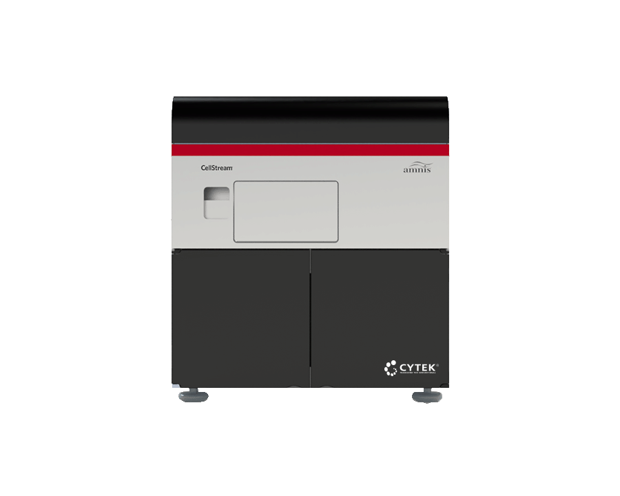
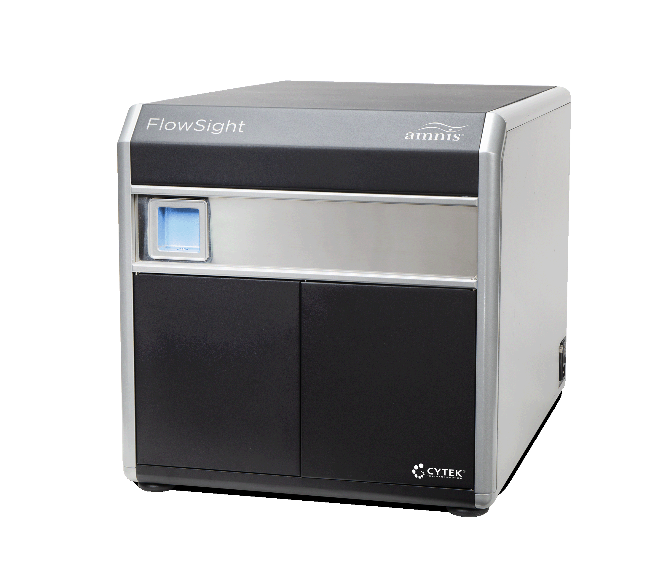
Cytek® Amnis® CellStream® | Cytek® Amnis® FlowSight® | Cytek® Amnis® ImageStream®X MKII |
| Attribute | Sensitive | Visual | Boundless |
| Lasers | 1-7 | 1-4 | 1-6 |
| Detection Type | CCD-TDI | CCD-TDI | CCD-TDI |
| Intensity | Up to 22 | Up to 10 | Up to 10 |
| Morphology | 3 | >1,000 | >1,000 |
| Magnification | N/A | 20x | Up to 60x |
| Format | Open | Open / Pre-Optimized Kits | Open / Pre-Optimized Kits |
| Flexibility | Highest | High | Highest |
| Microscopy Applications | Event Gallery | Yes | Yes |
| Learn More | Learn More | Learn More | |

ImageStream®X Mk II Optical Layout
Cells are fluorescently labeled in suspension and then loaded into the system using a syringe pump. Samples are hydrodynamically focused in a flow stream, and fluorescence is excited using multiple laser lines. The brightfield, SSC, and fluorescent images are captured by a 20x, 40x, or 60x imaging objective and transferred to the filter stack where each color is projected to a spatially discrete channel on the cameras. Up to 10 colors of fluorescence are collected in spatial registry, and each probe is measured for intensity, cellular morphology, and relative location to other probes.
CellStream® Optical Layout
Our patented time delay integration (TDI) and camera technology deliver sensitivity and expandability beyond what is possible with traditional flow cytometers:
- Up to 7 lasers are focused in discrete locations, aligned to four zones.
- Syringe Driven Fluidics: Hydrodynamically focused cells pass through the laser-illuminated region. Fluorochromes bound to the cells are excited and emit into the collection system. Fluorescence is collected and directed toward an intermediate image plane.
- A six-channel filter stack decomposes each of the four discrete vertical positions in the intermediate image plane into 22 separate channels of data.
- All 22 channels fit efficiently onto a charge-coupled device (CCD) array. The sensor contains multiple discrete collection fields using the same CCD as patented Amnis® technology.

For Research Use Only. Not for use in diagnostic or therapeutic procedures.