


CD7 Monoclonal Antibody (eBio124-1D1 (124-1D1)), Super Bright™ 436, eBioscience™
View Cart or Continue shopping.
Description
PRODUCT DETAILS
Host: Mouse
Isotype: IgG1, kappa
Clonality: Monoclonal
Clone: eBio124-1D1 (124-1D1)
Format: Super Bright™ 436
Reactivity: Human
Application: Flow Cytometry
Tested Dilution: 5 µL (0.25 µg)/test
Concentration: 5 µL/Test
Storage: 4° C, store in dark, DO NOT FREEZE!
Formulation: PBS, pH 7.2, containing 0.09% sodium azide
Purification: Affinity chromatography
Data Sheet: TDS
Specific Information
Description: The eBio124-1D1 monoclonal antibody reacts with human CD7, also known as gp40 and Leu9. CD7, a 40 kD receptor, is a member of the immunoglobulin gene superfamily. The N-terminal amino acid sequence (aa1-107) is highly homologous to Ig kappa light chain sequence; while the carboxyl-terminal region of the extracellular domain is proline-rich and has been postulated to form a stalk from which the Ig domain projects. CD7 is expressed on the majority of immature and mature T lymphocytes, and T cell leukemias. It is also found on natural killer cells, a small suppopulation of normal B cells and on maligant B cells. Cross-linking surface CD7 positively modulates T cell and NK cell activity, as measured by calcium flux, expression of adhesion molecules, cytokine secretion and proliferation. CD7 associates directly with phosphoinositol 3'-kinase. CD7 ligation induces production of D-3 phosphoinositides and tyrosine phosphorylation.
A clonogenic subpopulation of human CD34(+) CD38(-) cord blood cells that express CD45RA and HLA-DR and high levels of the CD7 has been reported. These cells possess the capacity for lymphopoiesis. They can generate B-cells, natural killer cells, and dendritic cells but do not possess the capacity to develop into myeloid cells or erythroid cells. The CD7(+) phenotype distinguishes primitive human lymphoid progenitors from pluripotent stem cells.
Furthermore, it has been suggested that CD7 co-operates with CD28 during Treg function, as mice deficient in both CD28 and CD7 have reduced total numbers of Tregs and these Tregs have reduced suppressive activity.
Applications Reported: This eBio124-1D1 antibody has been reported for use in flow cytometric analysis.
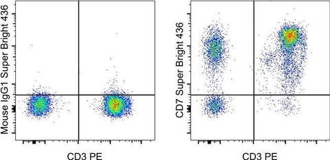
Applications Tested: This eBio124-1D1 antibody has been pre-diluted and tested by flow cytometric analysis of normal human peripheral blood cells. This may be used at 5 µL (0.25 µg) per test. A test is defined as the amount (µg) of antibody that will stain a cell sample in a final volume of 100 µL. Cell number should be determined empirically but can range from 10^5 to 10^8 cells/test.
Super Bright 436 can be excited with the violet laser line (405 nm) and emits at 436 nm. We recommend using a 450/50 bandpass filter, or equivalent. Please make sure that your instrument is capable of detecting this fluorochrome.
When using two or more Super Bright dye-conjugated antibodies in a staining panel, it is recommended to use Super Bright Complete Staining Buffer (Product # SB-4401) to minimize any non-specific polymer interactions. Please refer to the datasheet for Super Bright Staining Buffer for more information.
Fixation: Samples can be stored in IC Fixation Buffer (Product # 00-8222) (100 µL of cell sample + 100 µL of IC Fixation Buffer) or 1-step Fix/Lyse Solution (Product # 00-5333) for up to 3 days in the dark at 4°C with minimal impact on brightness and FRET efficiency/compensation. Some generalizations regarding fluorophore performance after fixation can be made, but clone specific performance should be determined empirically.
Excitation: 405 nm; Emission: 436 nm; Laser: Violet Laser
Super Bright Polymer Dyes are sold under license from Becton, Dickinson and Company.
For Research Use Only. Not for use in diagnostic procedures. Not for resale without express authorization.