


CD54 (ICAM-1) Monoclonal Antibody (eBioKAT-1 (KAT-1, KAT1)), Super Bright™ 436, eBioscience™
View Cart or Continue shopping.
Description
PRODUCT DETAILS
Host: Rat
Isotype: IgG2a, kappa
Clonality: Monoclonal
Clone: eBioKAT-1 (KAT-1, KAT1)
Format: Super Bright™ 436
Reactivity: Mouse
Application: Flow Cytometry
Tested Dilution: 0.5 µg/test
Concentration: 0.2 mg/mL
Storage: 4° C, store in dark, DO NOT FREEZE!
Formulation: PBS, pH 7.2, containing 0.09% sodium azide
Purification: Affinity chromatography
Data Sheet: TDS
Specific Information
Description: The eBioKAT-1 monoclonal antibody reacts with mouse CD54 (ICAM-1), which is a 95 kDa member of the immunoglobulin superfamily. CD54 is expressed at low levels on leukocytes and high endothelial venules, and expression increases in response to inflammatory cytokines. ICAM-1 binds to LFA-1 and this interaction is required for the transendothelial migration of T cells. ICAM-1-deficient mice are viable, however the migration of leukocytes to sites of inflammation is reduced leading to impaired immune and inflammatory responses. Based on the regulated expression of ICAM-1, it has been suggested that ICAM-1 may increase leukocyte extravasation at sites of inflammation, whereas the constitutively high expression of ICAM-2 mediates leukocyte traffic into non-inflamed tissue. The eBioKAT-1 monoclonal antibody recognizes a different epitope than the YN1/1.7.4 monoclonal antibody.
Applications Reported: This eBioKAT-1 antibody has been reported for use in flow cytometric analysis.
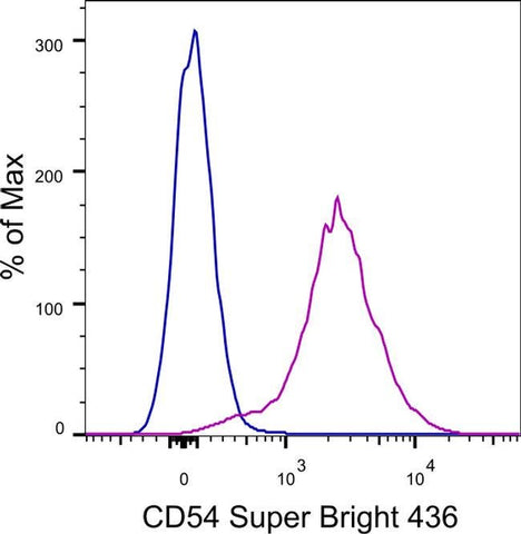
Applications Tested: This eBioKAT-1 antibody has been tested by flow cytometric analysis of mouse splenocytes. This may be used at less than or equal to 0.5 µg per test. A test is defined as the amount (µg) of antibody that will stain a cell sample in a final volume of 100 µL. Cell number should be determined empirically but can range from 10^5 to 10^8 cells/test. It is recommended that the antibody be carefully titrated for optimal performance in the assay of interest.
Super Bright 436 can be excited with the violet laser line (405 nm) and emits at 436 nm. We recommend using a 450/50 bandpass filter, or equivalent. Please make sure that your instrument is capable of detecting this fluorochrome.
When using two or more Super Bright dye-conjugated antibodies in a staining panel, it is recommended to use Super Bright Complete Staining Buffer (Product # SB-4401) to minimize any non-specific polymer interactions. Please refer to the datasheet for Super Bright Staining Buffer for more information.
Excitation: 405 nm; Emission: 436 nm; Laser: Violet Laser
Super Bright Polymer Dyes are sold under license from Becton, Dickinson and Company.
For Research Use Only. Not for use in diagnostic procedures. Not for resale without express authorization.