


CD185 (CXCR5) Monoclonal Antibody (MU5UBEE), Super Bright™ 436, eBioscience™
View Cart or Continue shopping.
Description
PRODUCT DETAILS
Host: Mouse
Isotype: IgG2b, kappa
Clonality: Monoclonal
Clone: MU5UBEE
Format: Super Bright™ 436
Reactivity: Human, Non-human primate, Rhesus Monkey
Application: Flow Cytometry
Tested Dilution: 5 µL (1.0 µg)/test
Concentration: 5 µL/Test
Storage: 4° C, store in dark, DO NOT FREEZE!
Formulation: PBS, pH 7.2, containing 0.09% sodium azide
Purification: Affinity chromatography
Data Sheet: TDS
Specific Information
Description: The MU5UBEE monoclonal antibody reacts with human and non-human primate CD185. CD185, which is also known as C-X-C chemokine receptor 5 (CXCR5) and Burkitt lymphoma receptor 1 (BLR1), is a seven transmembrane G protein-coupled receptor originally identified in Burkitt's lymphoma. In peripheral blood, CD185 is expressed on B cells, CD4+ T cells (but not Th1 or Th2 cells), as well as on a subpopulation of memory (CD45RO+) T cells. Circulating CD185+ T cells are in a resting state and migrate to the lymph nodes due to expression of CCR7 and CD62L. In tonsil, CD185 is expressed on nearly all CD4+ cells together with CD45RO and activation markers such as CD69 and ICOS. Tonsillar CD185+ cells have been shown to induce antibody production when co-cultured with B cells, thus supporting their role in providing help to B cells. Furthermore, this chemokine receptor plays a critical role in lymphocyte trafficking, in particular T cell migration into the B cell follicles of germinal centers in response to CXCL13, making CD185 an established marker of follicular helper T cells.
Applications Reported: This MU5UBEE antibody has been reported for use in flow cytometric analysis.
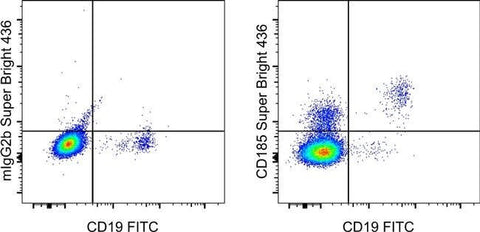
Applications Tested: This MU5UBEE antibody has been pre-diluted and tested by flow cytometric analysis of normal human peripheral blood cells. This may be used at 5 µL (1.0 µg) per test. A test is defined as the amount (µg) of antibody that will stain a cell sample in a final volume of 100 µL. Cell number should be determined empirically but can range from 10^5 to 10^8 cells/test.
Super Bright 436 can be excited with the violet laser line (405 nm) and emits at 436 nm. We recommend using a 450/50 bandpass filter, or equivalent. Please make sure that your instrument is capable of detecting this fluorochrome.
When using two or more Super Bright dye-conjugated antibodies in a staining panel, it is recommended to use Super Bright Complete Staining Buffer (Product # SB-4401) to minimize any non-specific polymer interactions. Please refer to the datasheet for Super Bright Staining Buffer for more information.
Excitation: 405 nm; Emission: 436 nm; Laser: Violet Laser
Super Bright Polymer Dyes are sold under license from Becton, Dickinson and Company.
For Research Use Only. Not for use in diagnostic procedures. Not for resale without express authorization.