


CD133 (Prominin-1) Monoclonal Antibody (TMP4), Super Bright™ 436, eBioscience™
View Cart or Continue shopping.
Description
PRODUCT DETAILS
Host: Mouse
Isotype: IgG1, kappa
Clonality: Monoclonal
Clone: TMP4
Format: Super Bright™ 436
Reactivity: Human
Application: Flow Cytometry
Tested Dilution: 5 µL (0.25 µg)/test
Concentration: 5 µL/Test
Storage: 4° C, store in dark, DO NOT FREEZE!
Formulation: PBS, pH 7.2, containing 0.09% sodium azide
Purification: Affinity chromatography
Data Sheet: TDS
Specific Information
Description: The TMP4 monoclonal antibody reacts with human CD133 (Prominin-1), a 120 kDa member of the pentaspan family of proteins, which also includes Prominin-2. Their expression is found within plasma membrane protrusions such as epithelial microvilli. CD133 can exist in a number of alternatively spliced isoforms, and the protein has several N-linked glycosylation sites: the occurrence of both may be tissue-dependent. Human CD133 was first identified as an epitope expressed on CD34+ hematopoietic progenitors. Although the ligand and function of CD133 remain unknown, it has since proven to be very useful as a marker for both stem cells and cancer stem cells. In addition to its expression on hematopoietic precursors, CD133 has been used to identify tumorigenic colon cancer stem cells, brain cancer stem cells, prostate cancer stem cells, in addition to others.
The binding of the TMP4 antibody does not block the binding of another anti-human CD133 antibody, EMK08 (cat. 12-1339) indicating that they recognize distinct epitopes.
Applications Reported: This TMP4 antibody has been reported for use in flow cytometric analysis.
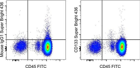
Applications Tested: This TMP4 antibody has been pre-diluted and tested by flow cytometric analysis of normal human peripheral blood cells. This may be used at 5 µL (0.25 µg) per test. A test is defined as the amount (µg) of antibody that will stain a cell sample in a final volume of 100 µL. Cell number should be determined empirically but can range from 10^5 to 10^8 cells/test.
Super Bright 436 can be excited with the violet laser line (405 nm) and emits at 436 nm. We recommend using a 450/50 bandpass filter, or equivalent. Please make sure that your instrument is capable of detecting this fluorochrome.
When using two or more Super Bright dye-conjugated antibodies in a staining panel, it is recommended to use Super Bright Complete Staining Buffer (Product # SB-4401) to minimize any non-specific polymer interactions. Please refer to the datasheet for Super Bright Staining Buffer for more information.
Excitation: 405 nm; Emission: 436 nm; Laser: Violet Laser
Super Bright Polymer Dyes are sold under license from Becton, Dickinson and Company.
For Research Use Only. Not for use in diagnostic procedures. Not for resale without express authorization.