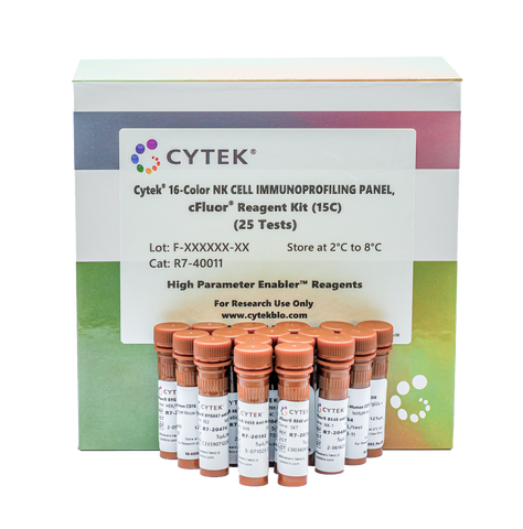
Cytek® 16-Color NK Cell Immunoprofiling Panel
View Cart or Continue shopping.
Description
The Cytek® 16-Color NK Cell Immunoprofiling Panel is designed and optimized by Cytek scientists to identify the three major human NK cell subsets, define the functional status within the three major subsets and identify additional NK cell subsets.
This 18-marker, 16-Color Human NK Cell Immunoprofiling Panel is comprised of the Cytek® 16-Color NK Cell Immunoprofiling Panel, cFluor® Reagent Kit (15C) (P/N: R7-40011) and the CD69 Monoclonal Antibody (FN50), Super Bright™ 436, eBioscience™ (P/N: 62-0699-42). The panel has been optimized for analyzing human PBMCs and whole blood on Cytek’s Northern Lights™ or Cytek Aurora™ systems equipped with violet, blue, and red lasers.
The Cytek® 16-Color NK Cell Immunoprofiling Panel enables the identification and characterization of the three major human NK cell subsets1,2 (CD16-CD56bright, CD16+CD56dim and CD16+CD56-) in human PBMCs and whole blood. The reagents in this kit also support analysis of the maturation and activation status, migratory potential, and the presence of NK activating and inhibitory receptors on the three major human NK cell subsets as well as identifying additional subsets. Innate Lymphoid Cells (ILCs) can be distinguished from NK cells by excluding the CD127+HLA-DR- population or characterized by analyzing the CD127+HLA-DR- population.
Tested Dilution: 5 μL / test
Application: Flow Cytometry
Storage: 2-8°C and protected from Light. (Do not freeze)
Formulation: Phosphate-buffered solution, pH 7.2, containing 0.09% sodium azide and 0.2% BSA.
DATA
Figure 1: Data analysis gating strategy for identifying NK cells and their activation status using the Cytek® 16-Color NK Cell Immunoprofiling Panel. Data were generated using whole blood collected in an EDTA tube from a healthy donor. After sequential gating of singlets and non-red blood cells (non-RBCs), NK cells are defined by sequential gating on lineage (CD14, CD19, CD123) negative, CD3-, lymphocytes, and HLA-DR-CD127-events. CD56 is plotted vs. CD16 to define the three major NK cell subsets in the periphery: CD16-CD56bright (Early NK cells), CD16+CD56dim (Mature NK cells), and CD16+CD56- (Terminal NK cells). Each NK cell marker is displayed vs. CD56, as well as CD159a vs CD159c, in density plots representing the total NK cell population.
The panel contains the following single vial reagents.
Table 1: Cytek® 16-Color NK Cell Immunoprofiling Panel (P/N R7-40011) composition.
RECOMMENDED USAGE
Human fresh whole blood (EDTA) and frozen PBMC samples have been tested to validate the performance of this kit. Please refer to the product web page for the staining protocols, fluorochrome list, experiment template and data acquisition protocol.
Please briefly centrifuge the reagent vial before use.
Use appropriate personal protective equipment per the product safety data sheet when using this product.
- Download the Sample Preparation (PBMC) Guidelines for the Cytek® 16-Color NK Cell Immunoprofiling Panel
- Download the Sample Preparation (Whole Blood) Guidelines for the Cytek® 16-Color NK Cell Immunoprofiling Panel
- Download the Acquisition Protocol for the Cytek® 16-Color NK Cell Immunoprofiling Panel
- Download the Cytek® 16-Color NK Cell Immunoprofiling Panel PBMC Template
- Download the Cytek® 16-Color NK Cell Immunoprofiling Panel WB Template
REFERENCES
- Poli A, et al. Immunology 126(4), 458 (2009)
- Cichocki F, et al. Front. Immunol. 10, 2078 (2019)
- Wei HY, et al. Sci. Rep. 6, 34310 (2016)
- van de Winkel J, et al. Today. 14, 215 (1993)
- Wright SD, et al. Science 249, 1431(1990)
- Ziegler-Heitbrock HWL, et al. Immunol. Today 14(3), 121 (1993)
- Angel CE, et al., J. Immunol. 176(10), 2006
- Wang K, et al. Exp. Hematol. Oncol. 1, 36 (2012)
- Bradbury LE, et al. J. Immunol. 151, 2915 (1993)
- Moretti S, et al. J. Biol. Regul. Homeost. Agents 15(1), 98 (2001)
- Schackelford DA, et al. Immunol. Rev. 66, 133 (1982)
- Van Dongen JJ, et al. Blood 71(3), 603
- Goldsmith MA, et al. Adv. Exp. Med. Biol. 234, 195 (1988)
- Lanier LL, et al. Fed. Proc. 45(12), 2823 (1986)
- Mason D, et al. Leukoc. Biol.211, 685 (2001)
- Stannard KA, et al. Blood Advances 3(11), 1681 (2019)
- Huang, et al. Cell Dev. Biol. 8, 564 (2020)
- Shibuya K, et al. Exp. Med.198, 1829 (2003)
- Dongchu M, et al. J. Haematol. 74,228 (2005)
- Martinet L, et al. Rev. Immunol.15, 243 (2015)
- Balsamo M, et al. Hematol.37, 1167 (2009)
- Lozano E, et al. Immunol.188, 3869 (2012)
- Tilden AB, et al. Nat. Immun. Cell Growth Regul. 5(2), 90 (1986)
- Chattopadhyay PK, et al. J. Leukoc. Biol. 85(1), 88 (2009)
- Lopez-Vergès S, et al. Blood 116(19), 3865 (2010)
- Kared H, et al. Cancer Immunol. Immunother. 65(4), 441 (2016)
- Houchins JP, et al. J. Exp. Med. 173(4), 1017 (1991)
- López-Botet M, et al. Immunol. 148(3), 155 (1997)
- Houchins JP, et al. J. Immunol. 158(8), 3603 (1997)
- Pende D, et al. J. Exp. Med. 190(10), 1505 (1999)
- Correia DV, et al. 118, 992 (2011)
- Sivori S, et al. Exp. Med.186(7), 1129 (1997)
- Hadad H, et al. Front. Immunol. 6, 495 (2015)
- Narni-Mancinelli E, et al. Natl. Acad. Sci. 108(45), 18324 (2011)
- Cibrián D, et al. Eur. J. Immunol. 47(6), 946 (2017)
- Yao X, et al. Cytokine Growth Factor Rev. 59, 37 (2021)
- Mueller A, et al. Int. J. Biochem. Cell Biol. 36, 35 (2004)
- Wang X, et al. Front. Immunol. 13, 960852 (2022)
- Clark MR, et al. Nat. Rev. Immunol. 14(2), 69 (2014) -CD127
- Björkström NK, et al. Immunology 139(4), 416 (2013) -CD127
- Surh CD, et al. Immunity 29(6), 848 (2008) -CD127
- Liu W, et al. J. Exp. Med. 203(7), 1701 (2006)-CD127
- Carapito R, et al. Immunol. Rev. 267(1), 88 (2015)
- Kurioka A, et al. Front. Immunol. 9(9), 486 (2018)
- Fergusson JR, et al. Cell Rep. 9(3), 1075 (2014)
- Van Acker HH, et al. Immunol. 8, 892 (2017)
- Crossland DL, et al. Oncogene. 37, 3686 (2018)
- Seidenfaden R, et al. Neurochem. Int. 49, 1 (2006)
- Barrow AD, et al. Front. Immunol. 10, 909 (2019)
For Research Use Only. Not intended for use in diagnostic procedures.
High Parameter Enabler™ Reagents
cFluor® B515, cFluor® 548, cFluor BYG610®, and cFluor® R720 are equivalent to CF®488A, CF®514, PE-CF®596R, and CF®700 respectively, manufactured and provided by Biotium, Inc. under an Agreement between Biotium and Cytek (LICENSEE). The manufacture, use, sale, offer for sale, or import of the product is covered by one or more of the patents or pending applications owned or licensed by Biotium. The purchase of this product includes a limited, non-transferable immunity from suit under the foregoing patent claims for using only this amount of product for the purchaser’s own internal research. No right under any other patent claim, no right to perform any patented method, and no right to perform commercial services of any kind, including without limitation reporting the results of purchaser’s activities for a fee or other commercial consideration, is conveyed expressly, by implication, or by estoppel.
cFluor® BYG667, cFluor® BYG710, and cFluor® BYG781 are tandem dyes made with R-PE. cFluor® R840 is a tandem dye made with APC. Caution – Tandem dyes may show changes in their emission spectra with prolonged exposure to light or fixatives.


