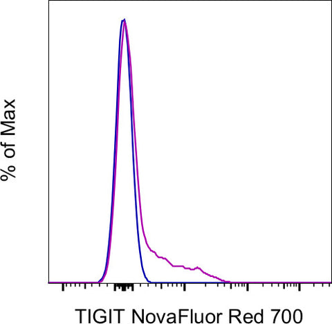
TIGIT Monoclonal Antibody (MBSA43), NovaFluor™ Red 700
カートを見る または ショッピングを続ける
説明
The MBSA43 monoclonal antibody reacts with human T cell immunoreceptor with Ig and ITIM domains (TIGIT), which is also known as VSTM3 or WUCAM. This 26 kDa protein is a member of the poliovirus receptor (PVR) family. The expression of TIGIT has been reported on NK cells, regulatory T cells, follicular T helper cells, memory CD4+ T cells, and CD8+ T cells, but it is not expressed on B cells or naive CD4+ T cells. TIGIT may be upregulated on naive CD4+ T cells upon activation. TIGIT can interact with certain members of the PVR and PVR-like families, including CD155, and also mediates the interaction of NK and T cells with antigen presenting cells, fibroblasts, and endothelial cells that express PVR and PVR-like proteins.
MBSA43 is reported to crossreact with Rhesus macaque.
Applications Reported: This MBSA43 antibody has been reported for use in flow cytometric analysis.
Applications Tested: This MBSA43 antibody has been pre-titrated and tested by flow cytometric analysis of normal human peripheral blood cells. This can be used at 5 µL (0.8 µg) per test. A test is defined as the amount (µg) of antibody that will stain a cell sample in a final volume of 100 µL. Cell number should be determined empirically but can range from 10^5 to 10^8 cells/test.
Each NovaFluor conjugate or kit is shipped with CellBlox Blocking Buffer. Use this buffer whenever staining with NovaFluor conjugates, including single-color compensation controls using cells. Whenever possible, we recommend adding CellBlox Blocking Buffer to antibody cocktails/master mixes prior to combining with cells. Add 5 µL per sample (regardless of the number of NovaFluors in your panel) to use the antibody cocktail as intended. For single-color controls, use 5 µL of CellBlox Blocking Buffer per 100µL of cell sample containing 10^3 to 10^8 cells.
Excitation: 639 nm; Emission: 700 nm; Laser: 633-640 nm (Red) Laser
