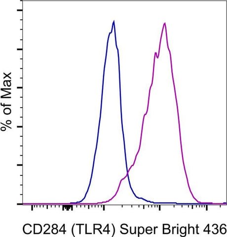
CD284 (TLR4) Monoclonal Antibody (HTA125), Super Bright™ 436, eBioscience™
カートを見る または ショッピングを続ける
説明
PRODUCT DETAILS
Host: Mouse
Isotype: IgG2a, kappa
Clonality: Monoclonal
Clone: HTA125
Format: Super Bright™ 436
Reactivity: Human
Application: Flow Cytometry
Tested Dilution: 5 µL (0.5 µg)/test
Concentration: 5 µL/Test
Storage: 4° C, store in dark, DO NOT FREEZE!
Formulation: PBS, pH 7.2, containing 0.09% sodium azide
Purification: Affinity chromatography
Data Sheet: TDS
Specific Information
Description: The HTA125 monoclonal antibody reacts with human Toll-like receptor 4 (TLR4). So far, at least ten members of the Toll family have been identified in humans. This family of type I transmembrane proteins is characterized by an extracellular domain with leucine-rich repeats and a cytoplasmic domain with homology to the type I IL-1 receptor. Two of these receptors, TLR2 and TLR4, are pattern recognition receptors and signaling molecules in response to bacterial lipoproteins and have been implicated in innate immunity and inflammation. TLR4 physically associates with another molecule called MD-2, and together with CD14, this complex is responsible for LPS recognition and signaling. TLR4 is expressed by peripheral blood monocytes. HTA125 has been reported to immunoprecipitate human TLR4 (~100 kDa) from transfected cells. Most TLR cell surface expression, especially TLR1 and TLR4, occurs at low levels on monocytes and at even lower levels on other cell types including granulocytes and immature dendritic cells (iDC). Furthermore, a relatively high degree of variability in TLR surface expression has been reported among normal donors.
It is highly recommended that for optimal staining of TLR4, whole blood be stained using the lysed whole blood protocol (found in Best Protocols) rather than Ficoll-gradient prepared normal human peripheral blood cells. The use of a density gradient appears to reduce the staining intensity significantly.
Applications Reported: This HTA125 antibody has been reported for use in flow cytometric analysis.
Applications Tested: This HTA125 antibody has been pre-diluted and tested by flow cytometric analysis of human TLR4/MD2-transfected cells. This may be used at 5 µL (0.5 µg) per test. A test is defined as the amount (µg) of antibody that will stain a cell sample in a final volume of 100 µL. Cell number should be determined empirically but can range from 10^5 to 10^8 cells/test.
Super Bright 436 can be excited with the violet laser line (405 nm) and emits at 436 nm. We recommend using a 450/50 bandpass filter, or equivalent. Please make sure that your instrument is capable of detecting this fluorochrome.
When using two or more Super Bright dye-conjugated antibodies in a staining panel, it is recommended to use Super Bright Complete Staining Buffer (Product # SB-4401-75) to minimize any non-specific polymer interactions. Please refer to the datasheet for Super Bright Staining Buffer for more information.
Excitation: 405 nm; Emission: 436 nm; Laser: Violet Laser
Super Bright Polymer Dyes are sold under license from Becton, Dickinson and Company.
For Research Use Only. Not for use in diagnostic procedures. Not for resale without express authorization.
